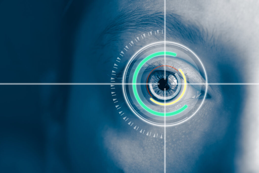
Cataract

What Do You Understand By Cataract?

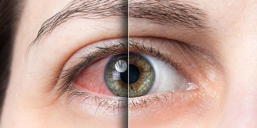
A cataract is a cloudiness or opacity of the eye’s natural clear lens. It obstructs the passage of light to the eye’s retina and impairs vision. When the lens develops cloudiness to the point that it impairs vision, it is called Cataract. It is like looking through a frosted glass.

What causes cataract?

Cataracts occur when there is a buildup of protein in the lens that makes it cloudy. This prevents light from passing clearly through the lens, causing some loss of vision. The common causes of cataract are:

  • Age related (most commonly)
  • Steroid use
  • Uveitis (inflammation)
  • Diabetes
  • Trauma
  • Congenital

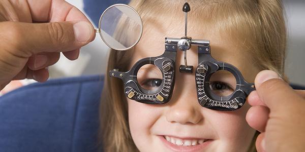
What’s the treatment for cataracts?

The only treatment of cataract is cataract surgery. There is NO medical treatment of cataract.

The cataract surgery involves removal of cataract by ultrasonic energy (phacoemulsification) and implantation of a permanent intraocular lens (IOL).

There have been many advances in the development of special intra-ocular lenses that correct for astigmatism and presbyopia in addition to myopia and hyperopia. These advances have allowed patients to strive for spectacle independence. There are a variety of lens options available; it is important to have a frank discussion with your surgeon to assess your visual needs and expectations.

Best cataract surgery in Pathankot

Saini Eye Hospital is the best hospital to get cataract surgery in Pathankot district with advanced technology and minimum risks involved.

High end Swiss made Phacoemulsification machine (Oertli) and Japanese operating microscope (Takagi) is used to ensure 100 % precision.

Our surgeons are trained at the best eye hospitals in Delhi and provide the latest and world class treatment options. MICS ( Micro Incision Cataract Surgery) is done in our hospital for all grades of cataracts. The surgery is completely painless and sutureless.

Lens implants

All types of lens implants are available to reduce the need for spectacles.

  • Traditional Monofocal IOL
  • EDOF (Extended Depth Of Focus) IOL
  • Multifocal IOL
  • Trifocal IOL
  • Toric IOL

Glaucoma

What is glaucoma?

Glaucoma or ‘Kala Motia’ is a condition wherein an increased intraocular pressure damages the optic nerve thereby affecting vision. A fluid known as aqueous nourishes the front part of the eye. In the normal eye, the rate of production of aqueous matches the rate of its drainage, thereby maintaining optimal pressure inside the eye.

Glaucoma is also called the silent thief of sight because in the early stages of the disease, there may be no symptoms. By the time glaucoma is detected, the patient has already suffered extensive peripheral vision damage which can no longer be restored. Early detection is the key to preserve vision. Once detected, glaucoma can be controlled by medicines (eye drops) or surgery to prevent further vision loss. Treatment of Glaucoma is life-long. Glaucoma treatment may include medical management, surgical management or management by lasers. The cause of glaucoma generally is a failure of the eye to maintain an appropriate balance between the amount of internal (intraocular) fluid produced and the amount that drains away.

The cause of glaucoma generally is a failure of the eye to maintain an appropriate balance between the amount of internal (intraocular) fluid produced and the amount that drains away.

Who Can Get it?

  • Anyone can get glaucoma. But along with other factors, you are at a higher risk of getting it if it runs in your family. If a close first degree relative had it, then you should fix an annual eye checkup without fail.
  • Diabetic/ Hypertensive individuals
  • Prolonged use of oral/topical corticosteroids
  • Following eye Trauma/inflammatory eye diseases such as uveitis.
  • Myopic eyes

What are the symptoms of glaucoma?

  • Seeing coloured halos around lights
  • Narrowing of vision (tunnel vision)
  • Pain & Redness in the eye
  • Hazy Cornea  ( in infants)
  • Nausea or vomiting, headache
  • Frequent change of eye glasses

There are two main types of glaucoma:

Open-angle glaucoma: Also called wide-angle glaucoma, this is the most common type of glaucoma. The structures of the eye appear normal, but fluid in the eye does not flow properly through the drain of the eye, called the trabecular meshwork.

Angle-closure glaucoma: Also called acute or chronic angle-closure or narrow-angle glaucoma, this type of glaucoma is less common in the West than in Asia. Poor drainage is caused because the angle between the iris and the cornea is too narrow and is physically blocked by the iris. This condition leads to a sudden buildup of pressure in the eye.

What’s the treatment for glaucoma?

Eye drops: When taken regularly and continuously as prescribed, eye drops control the eye pressure either by slowing the formation of aqueous fluid within the eye or by increasing the flow at the drainage area. The proper technique of using your medication is a key to successful treatment. Our glaucoma specialist or optometrist will teach you this technique.

Tablets: These are sometimes combined with the eye drops to decrease higher levels of eye pressure. They are occasionally known to cause tingling of fingers and toes, bowel irregularities, and in the very long term, kidney stones.

Laser surgery: Lasers are usually used in one of three ways:

  • In angle-closure glaucoma, the laser creates a hole in the iris to open up and improve the flow of aqueous fluid to the drain.
  • In painful late-stage glaucoma medication or surgery does not control the pressure. The laser closes some aqueous fluid-producing areas in the eye and lowers the eye pressure.

Operative Surgery (Trabeculectomy)

  • A new drainage channel is created to drain aqueous fluid from the eye.
  • Some cases may need Specialized Glaucoma Valve Surgery.
  • Surgery is recommended when our doctor feels that it is safer to operate than to allow optic nerve damage to continue.
  • The best treatment for you should be decided after a thorough examination and discussion with your specialist.

MIGS (Micro Invasive Glaucoma Surgery)

MIGS (Micro Invasive Glaucoma Surgery) is the latest cutting-edge technology to reduce intraocular pressure and the need for drops with a high safety profile and minimal disruption of ocular anatomy. It can be done combined with cataract surgery with a single incision and no need for sutures. The recovery is fast and operative risks are minimal. It is an ab-interno procedure.

Appropriate candidates for MIGS:

  • Open-angle Glaucoma
  • Patients on 2-3 anti-glaucoma eye drops undergoing cataract surgery

Patients with scarred conjunctiva MIGS devices:

  • KDB (Kahook Dual Blade)
  • I- Stent
  • BANG ( Bent Angle Needle Goniectomy)

Our glaucoma specialist Dr Tripti Johri is a trained Glaucoma Specialist from the best institute of north India – Dr Shroff’s Charity Eye Hospital, Delhi. She is well-versed in managing all kinds of complex glaucoma cases. She uses the latest technology, astute clinical acumen and empathetic behaviour to understand and treat her patients’ problems.

Vitreo Retina

Vitreoretinal Surgery

Saini Eye Hospital has the best retina specialist to provide medical and surgical management of all vitreo-retinal disorders like:

  • Diabetic Retinopathy
  • Vascular occlusions
  • Uveitis
  • ARMD ( Age Related Macular Degeneration)
  • ROP ( Retinopathy of prematurity)
  • Retinal Detachment
  • Ocular Foreign Body
  • Vitreous hemorrhage
  • CSR (Central Serous Retinopathy)

The facilities available at our hospital include:

  • OCT (Optical Coherence Tomography)

Topcon MAESTRO 2 OCT (Made in Japan) is available at our hospital for early and accurate diagnosis of retinal disorders.

  • MIVS (Micro Incision Vitrectomy Surgery)

MIVS is performed at our centre using the latest state-of-the-art vitrectomy machine using 25 gauge sutureless ports leading to excellent outcomes and fast recovery.

  • Intravitreal injections

All types of intravitreal injections are administered for diabetic retinopathy, ARMD, macular edema, CRVO, BRVO.

  • Retinal laser

High end ,Made in France,  QUANTEL laser is used to perform panretinal photocoagulation (PRP) and barrage laser.

  • ROP screening and treatment

Premature babies are screened for ROP and appropriate treatment is available in the form of injections and lasers.

  • Patient education and lifestyle counselling

Proper education is imparted to the patient to make lifestyle modifications to manage retinal diseases more effectively.

Cornea

Cornea Transplant Surgery & Treatment

Cornea is the outermost transparent layer of the eye which provides protection and accounts for approximately two-thirds of the eye’s total optical power. Our team at Saini Eye Hospital provides the best possible treatment for disorders of the cornea.

Corneal Ulcer

Corneal ulcer is a breach in the corneal layer along with infection. It can be bacterial , viral or fungal. The symptoms include eye redness, pain, decreased vision and discharge. Prompt diagnosis and best possible medical treatment is provided at our centre for all types of corneal ulcers. Corneal scraping and lab testing is done to reduce the infective load and accurately identify the organism responsible for the ulcer. Latest fortified antibiotics are started to give maximal medication. The causes of non-healing ulcers like high intraocular pressure, dry eye, associated adnexal infections, diabetes and immunocompromise are identified and treated concurrently.

Pterygium

Pterygium is an abnormal triangular growth of tissue on the conjunctiva (the clear membrane that covers the white of the eye) and the adjacent cornea (the clear front surface of the eye). It is associated with chronic sun and dust exposure. It causes redness, irritation, blurry vision and unsightly appearance.

Surgery is the only treatment with the preferred technique being LCAT (Limbal Conjunctival Autograft Transplantation) to minimise recurrence. This highly specialised surgery is performed at our centre which gives excellent outcomes.

Corneal Foreign Body

Impacted corneal foreign body is common in factory workers,  people who work with metal cutters, holi colours and firecracker injuries. Removal of corneal foreign body is done at our centre.

Chemical injury

Chemical injury to the cornea occurs commonly due to limestone (choona), industrial glue and firecrackers. It is an ocular emergency and should be treated immediately to save the eye and vision. Our team provides comprehensive treatment and on-call services to treat chemical injuries to the eye.

Paediatric Ophthalmologist

What is Pediatric Ophthalmology?

Pediatric ophthalmology is a specialized field that safeguards children’s vision and eye health. This subspecialty focuses on diagnosing and treating eye conditions in infants, children, and adolescents. Pediatric ophthalmologists are experts in managing childhood eye disorders, ensuring proper visual development and overall ocular health from birth through adolescence. Their expertise includes treating conditions like strabismus, amblyopia, congenital cataracts, and other vision impairments unique to younger patients.

Pediatric Ophthalmologist: Eye Care for Babies and Children

Our centre offers specialised Pediatric Ophthalmogy services to diagnose and manage pediatric eye disorders.

  • Refractive errors

Proper cycloplegic retinoscopy to measure the accurate number of eye glasses in children is very important for the visual development of children

  • Amblyopia ( Lazy Eye)

Diagnosis of underlying cause of amblyopia and its treatment is offered at our hospital. Amblyopia therapy, patching and nutritional guidance is done to achieve best possible outcomes.

  • Congenital cataract

Congenital cataract is treated using the latest machines and technology to restore vision in the child. Lens aspiration with posterior capsulotomy is performed to clear the visual axis so that the child can see clearly for life.

  • Congenital glaucoma

Congenital glaucoma is a serious vision threatening disease that can render the child blind if not treated promptly. We offer the best possible diagnosis and medical and surgical management of congenital glaucoma.

  • Retinopathy of prematurity (ROP)

Retinopathy of prematurity (ROP) is an eye disease in some premature babies born before 32 weeks. (A full-term pregnancy is about 38 to 42 weeks.) It is a problem that affects the tissue at the back of the eye called the retina. The retina senses light and sends signals to the brain so you can see. With ROP, unwanted blood vessels grow on the baby’s retina. These blood vessels can cause serious eye and vision problems later.

Risk factors for ROP:

  • Low birth weight ( under 3 kg or less).
  • How early a baby is born. A premature baby born at 28 weeks has a greater risk of having ROP than a premature baby born at 32 weeks.
  • Giving the infant extra oxygen after birth.

Shortly after birth, all premature babies should be checked for retinopathy of prematurity (ROP). An ophthalmologist can examine the infant’s eyes while they are in the hospital. However, ROP might not be visible until several weeks after birth. So, premature babies at risk for ROP are usually checked by an ophthalmologist at 4 to 6 weeks after birth and again thereafter.

It is important to take your baby to all follow-up appointments with your ophthalmologist. Timely detection and treatment can help prevent permanent vision loss.

At first, we monitor ROP to see if it goes away on its own. If abnormal blood vessels continue to grow, the infant’s eyes must be treated.

The ophthalmologist may treat ROP in one or more of the following ways:

  • Laser treatment
  • Freezing treatment (cryotherapy)
  • Medication eye injections(Anti-VEGF)
  • Surgery
  • Low birth weight ( under 3 kg or less).
  • How early a baby is born. A premature baby born at 28 weeks has a greater risk of having ROP than a premature baby born at 32 weeks.
  • Giving the infant extra oxygen after birth.

Shortly after birth, all premature babies should be checked for retinopathy of prematurity (ROP). An ophthalmologist can examine the infant’s eyes while they are in the hospital. However, ROP might not be visible until several weeks after birth. So, premature babies at risk for ROP are usually checked by an ophthalmologist at 4 to 6 weeks after birth and again thereafter.

It is important to take your baby to all follow-up appointments with your ophthalmologist. Timely detection and treatment can help prevent permanent vision loss.

At first, we monitor ROP to see if it goes away on its own. If abnormal blood vessels continue to grow, the infant’s eyes must be treated.

The ophthalmologist may treat ROP in one or more of the following ways:

Dry Eye

What is dry eye?

Dry eye is a condition in which a person doesn’t have enough quality tears to lubricate and nourish the eye. Tears are necessary for maintaining the health of the front surface of the eye and for providing clear vision.

Dry eye is a common and often chronic problem, particularly in older adults. With each blink of the eyelids, tears spread across the front surface of the eye, known as the cornea. Tears provide lubrication, reduce the risk of eye infection, wash away foreign matter in the eye and keep the surface of the eyes smooth and clear. Excess tears in the eyes flow into small drainage ducts in the inner corners of the eyelids, which drain into the back of the nose. Dry eyes can occur when tear production and drainage is not in balance.

What causes & risk factors for dry eye?

Dry eyes can occur when tear production and drainage are not in balance. People with dry eyes either do not produce enough tears or their tears are of a poor quality:

  • Inadequate amount of tears: Tears are produced by several glands in and around the eyelids. Tear production tends to diminish with age, with various medical conditions or as a side effect of certain medicines. Environmental conditions, such as wind and dry climates, can also decrease tear volume due to increased tear evaporation. When the normal amount of tear production decreases or tears evaporate too quickly from the eyes, symptoms of dry eye can develop.
  • Poor quality of tears:  Tears are made up of three layers: oil, water, and mucus. Each component protects and nourishes the front surface of the eye. A smooth oil layer helps prevent evaporation of the water layer, while the mucin layer spreads the tears evenly over the surface of the eye. If the tears evaporate too quickly or do not spread evenly over the cornea due to deficiencies with any of the three tear layers, dry eye symptoms can develop.

Dry eyes can develop for many reasons, including:

  • Age: Dry eyes are a part of the natural aging process. The majority of people over age 65 experience some symptoms of dry eyes.
  • Computer Vision Syndrome: This is a relatively new entity where excessive computer usage causes dry eye, headache, eye strain and blurry vision.
  • Gender: Women are more likely to develop dry eyes due to hormonal changes caused by pregnancy, the use of oral contraceptives and menopause.
  • Medications: Certain medicines, including antihistamines, decongestants, blood pressure medications, and antidepressants, can reduce tear production.
  • Medical conditions: People with rheumatoid arthritis, diabetes, and thyroid problems are more likely to have symptoms of dry eyes. Also, problems with inflammation of the eyelids (blepharitis), inflammationof the surfaces of the eye, or the inward or outward turning of eyelids can cause dry eyes to develop.
  • Environmental conditions: Exposure to smoke, wind and dry climates can increase tear evaporation resulting in dry eye symptoms. Failure to blink regularly, such as when staring at a computer screen for long periods of time, can also contribute to drying of the eyes.
  • Other factors: Long-term use of contact lenses can be a factor in the development of dry eyes. Refractive eye surgeries, such as LASIK, can decrease tear production and contribute to dry eyes.

Advanced dry eyes may damage the front surface of the eye and impair vision.

What are the symptoms of dry eye?

People with dry eyes may experience irritated, gritty, scratchy or burning eyes; a feeling of something in their eyes; excess watering; and blurred vision. Symptoms include:

  • stinging, scratching, or burning sensations.
  • Light Sensitivity.
  • Watery eyes.
  • Stringy mucus near the eye.
  • Blurry Vision

What’s the diagnosis for dry eye?

Dry eyes can be diagnosed through a comprehensive eye examination. Testing with emphasis on the evaluation of the quantity and quality of tears produced by the eyes may include:

  • Patient history to determine the patient’s symptoms and to note any general health problems, medications or environmental factors that may be contributing to the dry eye problem.
  • External examination of the eye, including lid structure and blink dynamics.
  • Evaluation of the eyelids and cornea using bright light and magnification.
  • Measurement of the quantity and quality of tears for any abnormalities. Special dyes may be put in the eyes to better observe tear flow and to highlight any changes to the outer surface of the eye caused by insufficient tears.

With the information obtained from testing, a doctor of optometry can determine if you have dry eyes and advise you on treatment options.

What’s the treatment for dry eye?

Treatments for dry eyes aim to restore or maintain the normal amount of tears in the eye to minimize dryness and related discomfort and to maintain eye health. Dry eyes can be a chronic condition, but a doctor of optometry can prescribe treatment to keep your eyes healthy and comfortable and to prevent your vision from being affected. The primary approaches used to manage and treat dry eyes include using over-the-counter artificial tear solutions, conserving tears, increasing tear production, and treating the inflammation of the eyelids or eye surface that contributes to the dry eyes.

  • Adding tears. Mild cases of dry eyes can often be managed using artificial tear solutions. These can be used as often as needed to supplement natural tear production. Preservative-free artificial tear solutions are recommended because they contain fewer additives, which can further irritate the eyes. People with dry eyes that don’t respond to artificial tears alone will need to take additional steps to treat their dry eyes.
  • Conserving tears. Keeping natural tears in the eyes longer can reduce the symptoms of dry eyes. This can be done by blocking the tear ducts through which the tears normally drain. The tear ducts can be blocked with tiny silicone or gel-like plugs that can be removed if needed. Or a surgical procedure can permanently close the tear ducts. In either case, the goal is to keep the available tears in the eye longer to reduce problems related to dry eyes.
  • Increasing tear production. A doctor of optometry can prescribe eye drops that increase tear production. Taking an omega-3 fatty acid nutritional supplement may also help.
  • Treating the contributing eyelid or ocular surface inflammation. A doctor of optometry might recommend prescription eye drops or ointments, warm compresses and lid massage, or eyelid cleaners to help decrease inflammation around the surface of the eyes.

What’s the prevention for dry eye?

You can take the following steps to reduce symptoms of dry eyes:

  • Remember to blink regularly when reading or staring at a computer screen for long periods of time.
  • While working on computers, take regular breaks using the 20-20-20rule: every 20 minutes, shift your eyes to look at an object at least 20 feet away, for at least 20 seconds.
  • Increase the humidity in the air at work and at home.
  • Wear sunglasses outdoors, particularly those with wraparound frames, to reduce exposure to drying winds and the sun.
  • Nutritional supplements containing essential fatty acids may help decrease dry eye symptoms in some people. Ask your doctor if taking dietary supplements could help your dry eye problems.
  • Avoiding becoming dehydrated by drinking plenty of water (8 to 10 glasses) each day.
  • Avoid air getting blown in your eyes by directing car heaters away from your face.
  • Avoid environments that are drier than normal, such as deserts, airplanes, and places at high altitudes.